|
Plate IV.Plate II
Click image to enlarge
 |
Ā |
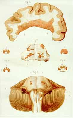
Plate IV. Charcot's Original Commentary:
Fig. 1, a-a, "Sclerosed plaque affecting the [upper] wall of the lateral
[cerebral] ventricle" - reaching, up to one centimeter, off the ventricular
border; fig. 2, a,a, "Sclerosed nuclei", exposed by sectioning the pons
parallel to its anterior front.
Fig. 3. a,a, "Sclerosed plaques of pons and spinal bulb"; b,b, "ependyma"
[membranous wall] of the brain's fourth ventricle.
Fig. 4, Spinal cord cross-sections (d, the cord's front) : A, above cervical
enlargement; B,B', at middle; C, three centimeters over lower end. The
spinal cord's lateral columns appeared throughout primarily and, in the lumbar
section, exclusively involved.
|
|
Specific Lesion Features:
Fig. 1 shows a massive, peripherally undulating epiventricular lesion
expanded into a cerebral hemisphere; it embeds the local venous blood vessels'
stems and their branches' proximal, i.e. more central, segments. Most
lesion-veins appear silhouetted by unevenly asymmetrically widened perivascular
spaces; the larger veins show a winding course and appear related to broader
lesion projections.
Fig. 2 shows winding and bumpy, multiheaded lesions expanded in the surface
(c.f. Plate II, fig. 1) and into the depth of the pons (c.f. fig. 3, a,a).
|
Figg. 3 and 4 confirm the specific involvement of, above all, the spinal
cord's flanks. The laterally based lesion wedges are partly interconnected
(figg. 4; B, B') and appear one times turned backwards (fig. 4: C, left side;
c.f. Plate I, fig. B, right side; Plate III, fig. 1'', left side).
Significance:
This plate, drawn by Charcot and presented in Ordenstein's 1867 Parisian
thesis, was the first to display specific ventricle-based lesion expansions
into the cerebral hemispheres. It forms the earliest and still unmatched
synopsis of the specific post-mortem findings of multiple sclerosis of brain
and spinal cord.
|