|
Plate VII
Click image to enlarge
 |
Ā |
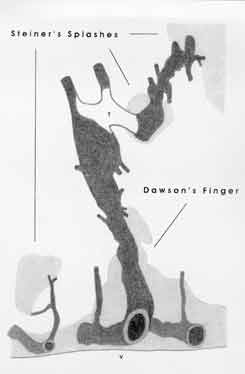
Plate IV.
Original Observations by Putnam and Adler:
"... lateral ventricles ... lined with gliotic tissue in which lay large
veins, many ... surrounded by hematogenous pigment. The sclerotic tissue
followed the radial veins for a variable distance, and irregular patches of
sclerosis were found at intervals along them ... a large dilated vein leading
toward the cortex was surrounded by a sleeve of plaque ... plaques surrounded
the larger (venous) trunks at intervals ... - Particularly striking was the
irregular, tortuous, congested contour of the main (strongest) (plaque-)
veins."
|
Lesion Specification:
The main lesion, indicated as a "Dawson's finger', forms roughly a cone which
rising off of the cerebral ventricular border (V), in a distinct relationship
to a strong vein stem. The latter, about one millimeter thick, empties (in the
midst of the figure's lower edge) into an epiventricular collecting vein. The
former lesion-vein shows, downstream to its thrombotic obstruction (T),
striking, peripherally increasing distensions: Its main branch exhibits,
downstream to the outermost "Steiner's splash", grotesque distortions. Two
other isolated plaques, or "Steiner's splashes", are seen to have emerged
immediately distant to the major lesion-vein's thrombus respectively to another
lesion-vein's peripheral trifurcation (veins in black, lesions in grey).
|
Documentary Significance:
This reconstruction model of Putnam and Adler's, of specific cerebral lesions
and their veins, offered the first concrete evidence of the lesion-veins'
peculiar deformities and of their apparently paradoxical spacial relationships
to not so much the ventricle based "Dawson's fingers" as to the cerebrum's
isolated plaques, or "Steiner's splashes".
|