|
Plate XIII.
Click image to enlarge
 |
Ā |
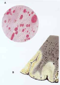
Oppenheim and Cassirer's Original 1907 Characterization of a
Subcortically Spread Multifocal Encephalitis:
Fig. 4, "Inflammatory foci" in cerebral cortex and subcortical white matter
at weak magnification; fig. 5, (hemorrhagic) white matter foci at stronger
magnification. The "dot-shaped" or streaky multifocal damages' perivascular
spread is obvious.
|
Lesion Character:
The illustrations of the dissemination of "encephalitic foci " according to a
random involvement of minor blood vessels, and of their evolution through
intensified effusions from transitional vessels make the differences between
"(blood-borne) encephalitis" and cerebral multiple sclerosis particularly
explicit.
|
Ā |
Significance of the Documentation:
The text to the given illustration pointed out that a larger "encephalitic
lesion's" origin in a confluence of small "inflammatory foci" may be evident
from an increasingly dense clustering of perivascular infiltrates towards a
corresponding larger lesion's edge. Large "encephalitic" lesions were thus
indicated to develop in a principally different way from the cerebral plaques
of multiple sclerosis.
|